Figure 5
Detection of Transferrin Oxidative Modification In vitro and In vivo by Mass Spectrometry. Hereditary Hemochromatosis is a Model
Mohamed Ahmed*
Published: 27 May, 2025 | Volume 9 - Issue 1 | Pages: 009-019
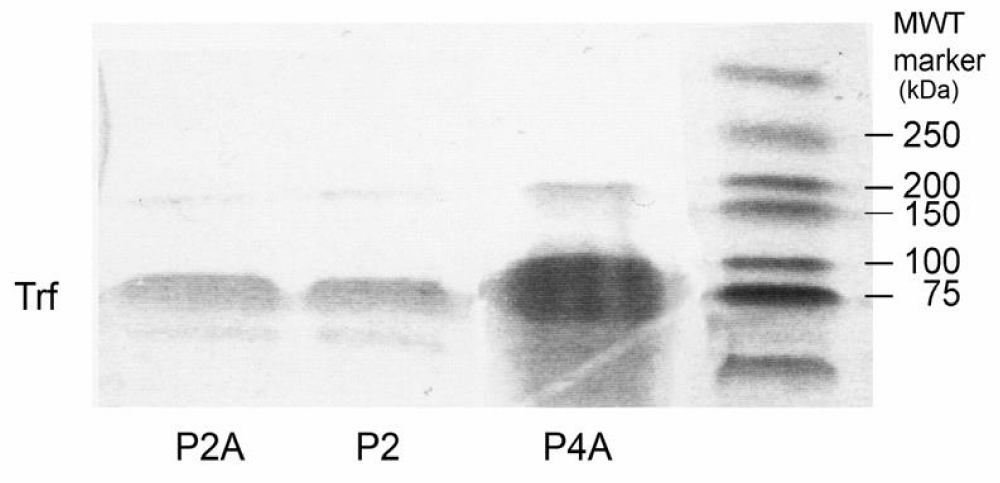
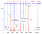
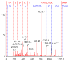
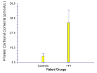
Figure 5:
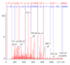
1-D SDS-PAGE shows transferrin bands (Trf), visualized by Coomassie stain at the molecular mass of 78 kDa from immunoprecipitants of plasma samples from patients with HH. The protein was loaded onto a 10% gel, stained by Coomassie, destained and digested into peptide fragments by trypsin.
Read Full Article HTML DOI: 10.29328/journal.apb.1001025 Cite this Article Read Full Article PDF
More Images
Similar Articles
-
The importance of gestational age in first trimester, maternal urine MALDI-Tof MS screening tests for Down SyndromeRay K Iles*,Nicolaides K,Pais RJ,Zmuidinaite R,Keshavarz S,Poon LCY,Butler SA. The importance of gestational age in first trimester, maternal urine MALDI-Tof MS screening tests for Down Syndrome. . 2019 doi: 10.29328/journal.apb.1001008; 3: 010-017
-
Detection of Transferrin Oxidative Modification In vitro and In vivo by Mass Spectrometry. Hereditary Hemochromatosis is a ModelMohamed Ahmed*. Detection of Transferrin Oxidative Modification In vitro and In vivo by Mass Spectrometry. Hereditary Hemochromatosis is a Model. . 2025 doi: 10.29328/journal.apb.1001025; 9: 009-019
Recently Viewed
-
Detrimental Effects of Methylenetetrahydrofolate Reductase (MTHFR) Gene Polymorphism on Human Reproductive Health: A ReviewVandana Rai*,Pradeep Kumar. Detrimental Effects of Methylenetetrahydrofolate Reductase (MTHFR) Gene Polymorphism on Human Reproductive Health: A Review. Clin J Obstet Gynecol. 2025: doi: 10.29328/journal.cjog.1001182; 8: 007-014
-
Fetal Bradycardia Caused by Maternal Hypothermia: A Case ReportMuna Alqralleh,Rahma Al-Omari,Shrouq Aldahabi,Doha Abdelbage,Maher Al-Hajjaj*,Lujain Alababesh. Fetal Bradycardia Caused by Maternal Hypothermia: A Case Report. Clin J Obstet Gynecol. 2025: doi: 10.29328/journal.cjog.1001180; 8: 001-002
-
Betty Neuman System Model: A Concept AnalysisAdnan Yaqoob*, Rafat Jan, Salma Rattani and Santosh Kumar. Betty Neuman System Model: A Concept Analysis. Insights Depress Anxiety. 2023: doi: 10.29328/journal.ida.1001036; 7: 011-015
-
Rare Locations of Plasma Cell Tumour: A Single-Centre ExperienceVladimir Prandjev,Donika Vezirska,Ivan Kindekov*. Rare Locations of Plasma Cell Tumour: A Single-Centre Experience. J Hematol Clin Res. 2025: doi: 10.29328/journal.jhcr.1001036; 9: 015-019
-
Developmentally appropriate practices on knowledge skills for contributing child’s intelligences of receptive language skills in appropriate and inappropriate early childhoodsNarida Rattana-Umpa,Jirawon Tanwatthanakul*,Churaporn Sota,Toansakul Tony Santiboon. Developmentally appropriate practices on knowledge skills for contributing child’s intelligences of receptive language skills in appropriate and inappropriate early childhoods. Arch Psychiatr Ment Health. 2021: doi: 10.29328/journal.apmh.1001034; 5: 042-050
Most Viewed
-
Feasibility study of magnetic sensing for detecting single-neuron action potentialsDenis Tonini,Kai Wu,Renata Saha,Jian-Ping Wang*. Feasibility study of magnetic sensing for detecting single-neuron action potentials. Ann Biomed Sci Eng. 2022 doi: 10.29328/journal.abse.1001018; 6: 019-029
-
Evaluation of In vitro and Ex vivo Models for Studying the Effectiveness of Vaginal Drug Systems in Controlling Microbe Infections: A Systematic ReviewMohammad Hossein Karami*, Majid Abdouss*, Mandana Karami. Evaluation of In vitro and Ex vivo Models for Studying the Effectiveness of Vaginal Drug Systems in Controlling Microbe Infections: A Systematic Review. Clin J Obstet Gynecol. 2023 doi: 10.29328/journal.cjog.1001151; 6: 201-215
-
Causal Link between Human Blood Metabolites and Asthma: An Investigation Using Mendelian RandomizationYong-Qing Zhu, Xiao-Yan Meng, Jing-Hua Yang*. Causal Link between Human Blood Metabolites and Asthma: An Investigation Using Mendelian Randomization. Arch Asthma Allergy Immunol. 2023 doi: 10.29328/journal.aaai.1001032; 7: 012-022
-
Impact of Latex Sensitization on Asthma and Rhinitis Progression: A Study at Abidjan-Cocody University Hospital - Côte d’Ivoire (Progression of Asthma and Rhinitis related to Latex Sensitization)Dasse Sery Romuald*, KL Siransy, N Koffi, RO Yeboah, EK Nguessan, HA Adou, VP Goran-Kouacou, AU Assi, JY Seri, S Moussa, D Oura, CL Memel, H Koya, E Atoukoula. Impact of Latex Sensitization on Asthma and Rhinitis Progression: A Study at Abidjan-Cocody University Hospital - Côte d’Ivoire (Progression of Asthma and Rhinitis related to Latex Sensitization). Arch Asthma Allergy Immunol. 2024 doi: 10.29328/journal.aaai.1001035; 8: 007-012
-
An algorithm to safely manage oral food challenge in an office-based setting for children with multiple food allergiesNathalie Cottel,Aïcha Dieme,Véronique Orcel,Yannick Chantran,Mélisande Bourgoin-Heck,Jocelyne Just. An algorithm to safely manage oral food challenge in an office-based setting for children with multiple food allergies. Arch Asthma Allergy Immunol. 2021 doi: 10.29328/journal.aaai.1001027; 5: 030-037

If you are already a member of our network and need to keep track of any developments regarding a question you have already submitted, click "take me to my Query."


